
Diagnostics
- Home
- Diagnostics
Dental diagnostics in Baku — 3D imaging, x-rays and digital scanning
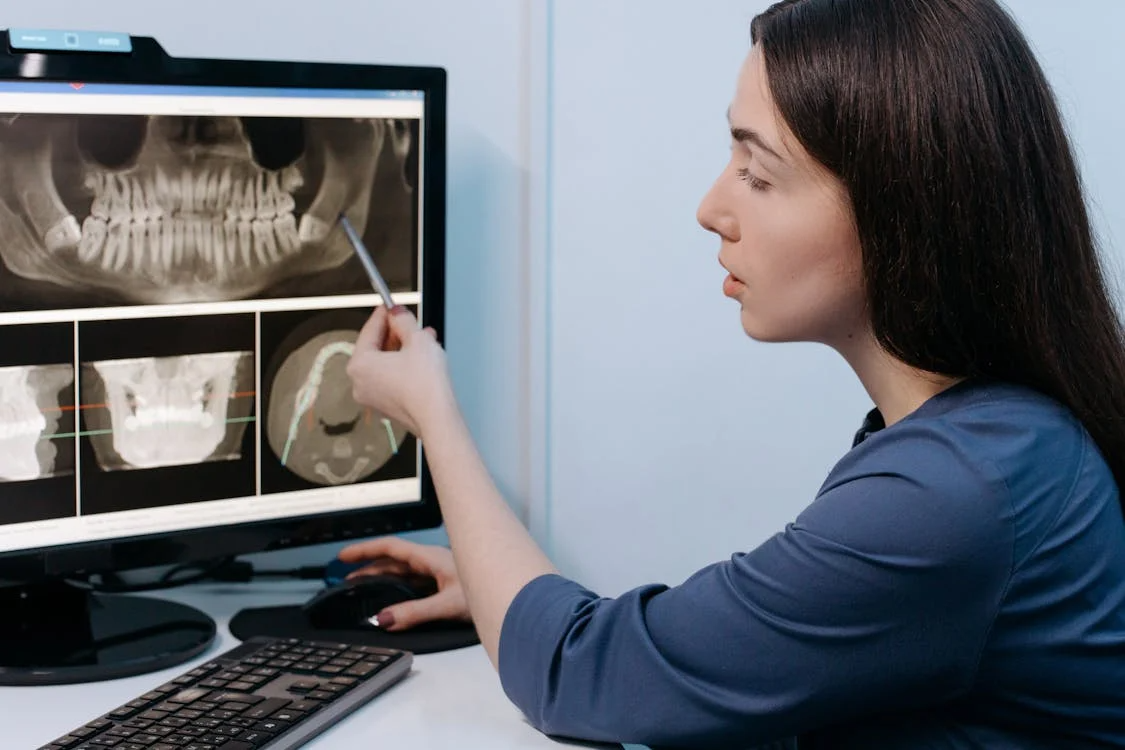
A treatment plan comes out of diagnostics: what is visible to the eye is only part of what is happening inside the tooth and in the bone. At German Dental Clinic diagnostics are digital — the result appears on screen straight away and is reviewed together with the patient.
What we use
- Digital radiography (RVG). A precise image of a single tooth and its root area — decay, canals, changes at the root tip.
- Panoramic x-ray (OPG). An overview of all the teeth, the jaws and the temporomandibular joint.
- CBCT / 3D imaging. Bone volume, the course of the nerve canals, the state of the sinuses — the basic study for implants and complex endodontics.
- Intraoral scanning. An impression taken without impression material; the model is built on screen.
- Photo protocol. For aesthetic planning and for comparing progress during treatment.
- Cephalometric analysis. Assessment of how the jaws relate to each other in orthodontics.
When diagnostics matter most
Before implant surgery — the position of an implant cannot be planned safely without knowing bone volume and nerve location. In root canal treatment — to find accessory and curved canals. In orthodontics — to assess jaw relationships and root positions. Before removing a wisdom tooth — to judge how close the root is to the nerve canal. And whenever the cause of pain is not obvious: a cyst, an abscess or a root fracture is often visible only on an image.
Is the radiation a concern
Digital sensors work at a markedly lower dose than film, and a single dental x-ray is comparable to a day of natural background radiation. The principle still holds: an image is taken only where indicated, pregnancy must be mentioned to the dentist, and protective equipment is used. CBCT gives more information than a panoramic x-ray and is ordered for a specific question — implant planning, for example.
What digital scanning is for
An intraoral scanner replaces conventional impression material: the procedure does not trigger a gag reflex, the result is immediately visible as a 3D model and goes to the laboratory electronically. That improves the fit of crowns, veneers, aligners and surgical guides and reduces the need for repeat impressions.
How the appointment runs
- Your complaint and medical history are recorded.
- Clinical examination — teeth, gums, bite and joint.
- An x-ray, CBCT or scan where indicated.
- The results are reviewed together on screen.
- A treatment plan: stages, order and indicative timelines.
What an image shows and what it does not
X-rays show hard tissue: the depth of decay, how well the canals are filled, changes at the root tip, bone level and cysts. They will not tell you whether the pulp is alive, what kind of pain you have or how inflamed the gum is — that needs a clinical examination, vitality testing and periodontal probing. A diagnosis is therefore never built on an image alone: the image confirms or refines what the examination found.
When a repeat image is needed
At routine check-ups control images are usually taken every 12–24 months, and more often where caries risk is high. After root canal treatment the result is checked with a control image within 6–12 months, and around implants bone level is monitored during the first year and at regular intervals afterwards. The exact schedule is always individual and set by the starting situation.
Imaging in children
A child is only imaged for a specific question: the state of a developing permanent tooth, trauma, a disturbed eruption sequence, or suspected decay between teeth that an examination cannot see. The dose is reduced for age, protective equipment is used, and the procedure is explained to the child in simple words beforehand — which helps them stay still and cuts the need for a repeat image.
Frequently asked questions
Can I use an x-ray from another clinic?
Usually yes, if it is recent enough and covers the area in question. Bring it as a file or on a disc and the dentist will say whether it needs repeating.
Can x-rays be taken during pregnancy?
In urgent situations yes, with a protective apron and a minimal dose. Routine imaging is normally deferred to the second trimester or until after the birth — tell the dentist about the pregnancy in advance.
How is CBCT different from a panoramic x-ray?
A panoramic x-ray is a two-dimensional overview. CBCT produces a three-dimensional image: bone thickness, the course of the nerves and how the structures sit relative to each other.
Can I take the results with me?
Yes, images are issued as files and can be shown to another specialist.
See also: Dental implants · Endodontics · Dental cyst · Dental abscess · What a gnathologist does
Book a diagnostic appointment — results are explained in the same visit and handed over together with the treatment plan.